|
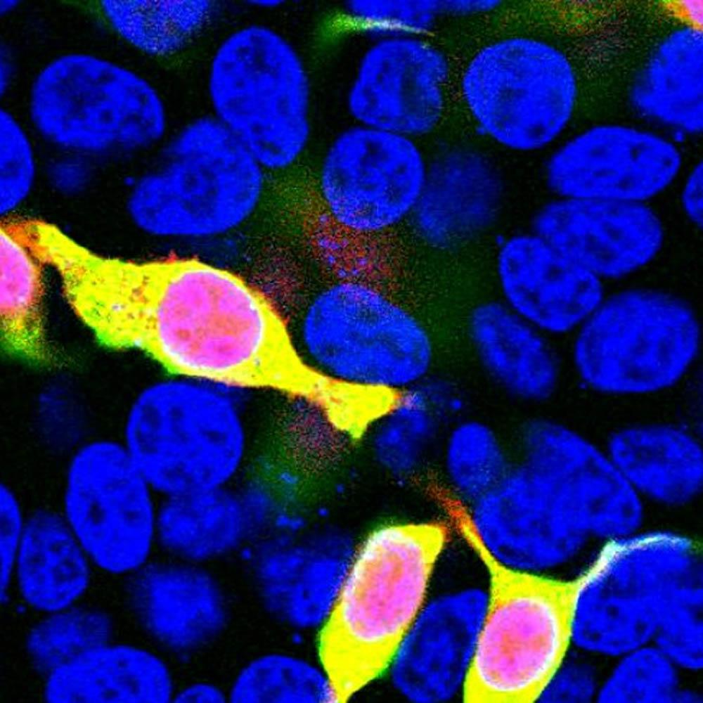
#M-1653-100: IF analysis of HEK293 cells transfected with mCherry protein construct, red, and stained mouse anti-mCherry antibody, M-1653-100, green. Transfected cells that overlap mCherry protein and anti-mCherry antibodies appear golden in color. Untransfected cells appear only to show their DAPI-stained blue nuclei.
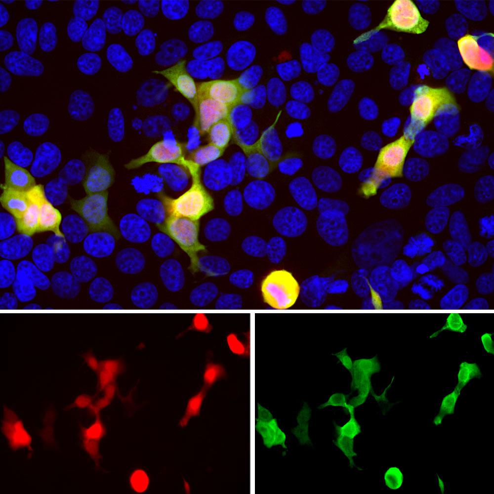
#C-1655-100:
IF: The confocal microscopical images show the HEK293 cells transfected with mCherry (red) and stained with C-1655-100 (green). The transfected mCherry (red) with chicken anti-mCherry antibody (green) together appear golden. Untransfected cells appear blue from a nuclei-only stain. The chicken anti-mCherry antibody provides a strong and specific signal.
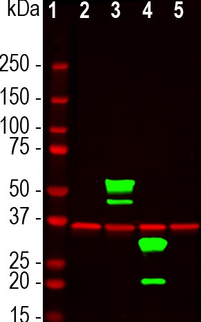
WB: It shows HEK293 mCherry expressing cell lysates using chicken pAb to mCherry,
C-1655-100, in green, and rabbit pAb to GAPDH, R-1701-100, in red: Lanes: [1] MW Markers, [2] untransfected HEK293 control cells, [3] HEK293 cells expressing two tdTomato protein domains, [4] HEK293 cells expressing mCherry-HA protein, and [5] HEK293 expressing GFP protein. C-1655-100 mCherry antibody recognizes tdTomato and mCherry proteins revealing major bands at about 60kDa and 30kDa but does not recognize GFP. The red band at 37kDa corresponds to GAPDH protein, used as a loading control.
#R-1654-100:
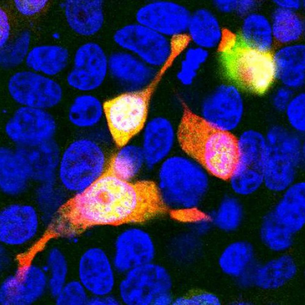
IF analysis of HEK293 cells transfected with mCherry protein construct, red, and stained with rabbit pAb to mCherry, R-1654-100, green. Nuclear DNA staining is blue. Red Green matching fluorescences of mCherry protein glow golden in this analysis, as expected. The R-1654-100 antibody shows only golden-colored transfected cells expressing mCherry protein and untransfected cells, which reveal only their nuclei.
|