|
Visualize LAMP1 localization with remarkable clarity and precision using
M-1690-100 antibody with various imaging techniques, such as Immunocytochemistry, Flow Cytometry, and Western Blotting.
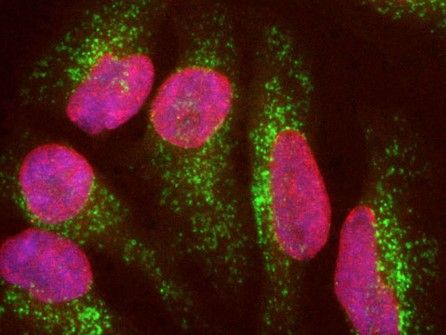
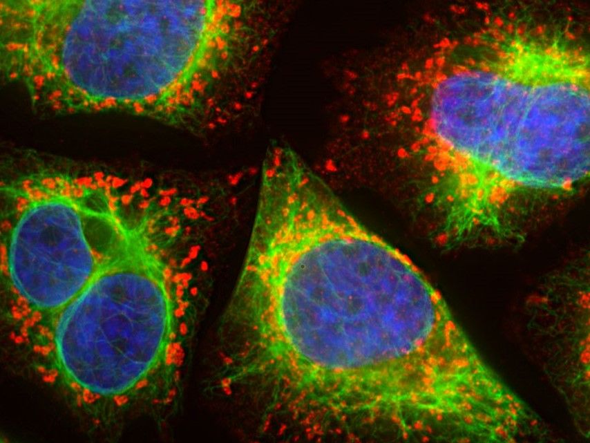
The image represents the LAMP1 antibody, which reveals the presence of vesicular LAMP1 protein within lysosomes. While the vimentin antibody imagines the intermediate filament network in these cells.
|