JOPA

 Journal of Orthopedics for Physician Assistants


Category 1 CME


April Image Quiz


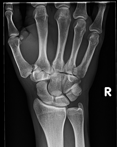
A 25-year-old man presents with right thumb pain after an all-terrain-vehicle accident 2 days ago. He had immediate pain and swelling after the injury, and he has been unable to use the thumb since. Radiographs obtained at an outside urgent-care facility are shown below.        


Figure 1. AP Wrist Radiograph   

Figure 2. Lateral Wrist Radiograph
 

Figure 3. Oblique Wrist Radiograph


What is the recommended treatment? 

A. Thumb spica cast immobilization
B. Activities to tolerance
C. Closed reduction and cast immobilization
D. Closed reduction and percutaneous pinning
E. Open reduction and internal fixation


The patient sustained a displaced Bennett fracture, which is a single vertical intra-articular fracture at the base of the carpometacarpal (CMC) joint. The most common mechanism of injury is a direct blow to the thumb with a partially flexed metacarpal. The strong volar oblique ligament, or palmar beak ligament, is the primary stabilizer of the trapeziometacarpal joint. The ligament holds the volar bone fragment in place as the main fragment of the metacarpal shaft displaces. The metacarpal shaft usually displaces radially and dorsally from the pulling forces of the abductor pollicis longus and the adductor pollicis. Subluxation of the CMC joint can be seen on the patient's radiographs. A Rolando fracture also involves the trapeziometacarpal joint but has a different fracture pattern with similar deforming forces. A Rolando fracture is Y-shaped with intra-articular comminution whereas a Bennett fracture has a single fracture fragment. Anteroposterior (AP), lateral, and oblique radiographs of the thumb are necessary to determine the type of fracture pattern present. Radiographs of a Bennett fracture will show an avulsion off the volar prominence of the metacarpal base as illustrated in Figure 1.


Figure 1. Bennett fracture.


Treatment of metacarpal base fractures is determined by the CMC joint stability, fracture pattern, and amount of displacement. Extra-articular fractures of the thumb metacarpal with angulation of up to 20 to 30 degrees in the lateral plane can be treated nonoperatively with a thumb spica cast for 6 weeks without functional deficit; however, the cosmetic appearance may be bothersome. Nondisplaced intra-articular fractures can also be treated conservatively but should be followed closely for displacement. Displaced intra-articular Bennett fractures treated nonoperatively lead to persistent subluxation and a likelihood of post-traumatic arthritis. Closed reduction should be attempted with the patient under anesthesia and use of fluoroscopic guidance to achieve a congruent joint space. The reduction maneuver includes longitudinal traction with abduction and extension of the thumb metacarpal. The amount of residual displacement after reduction correlates with the severity of arthritis, so an anatomic reduction is crucial. If closed reduction is achieved with <2 mm of displacement then percutaneous pinning is performed to stabilize the joint. The deforming forces at the CMC joint tend to displace the reduction, so percutaneous pinning of the metacarpal shaft to the trapezium is used to hold the metacarpal reduced. A pin is also placed through the fracture fragment and into the 2nd metacarpal base for further stability. Percutaneous pinning also provides sufficient stability for accurate healing of the stabilizing ligaments. The pins are removed approximately 6 weeks postoperatively. Open reduction and internal fixation is indicated when the fragment cannot be closed reduced with less than a 2-mm step-off and if the displaced fragment is >20% of the articular surface. Cast immobilization with the interphalangeal joint free is typically used for 6 to 8 weeks postoperatively. Progressive range-of-motion exercises are initiated after cast removal, with the goal of forceful pinch loading beginning at 3 months. Residual instability is a more prevalent complication than joint stiffness, so patients should be advised to adhere to a slowly progressive rehabilitation protocol. 

Answer D.


References

1. Soyer AD. Fractures of the base of the first metacarpal: current treatment options. J Am Acad Orthop Surg. 1999 Nov-Dec;7(6):403-12.

2. Henry MH. Fractures and dislocations of the hand. Bennett fracture. In: Bucholz RW, Heckman JD, Court-Brown C, editors. Rockwood and Green's fractures in adults. 6th ed. Philadelphia, PA: Lippincott Williams & Wilkins; 2005. p 836-850. 

3. Cullen JP, Parentis MA, Chinchilli VM, Pellegrini VD Jr. Simulated Bennett fracture treated with closed reduction and percutaneous pinning. A biomechanical analysis of residual incongruity of the joint. J Bone Joint Surg Am. 1997 Mar;79(3):413-20.


New on CME4PAC:  Bone Tumors Exam 
3 Category 1 SA Credits