High-specificity GAPDH Antibodies

Glyceraldehyde 3-Phosphate Dehydrogenase (GAPDH) is an important enzyme in the glycolytic pathway. It helps convert glucose into energy by catalyzing a specific step: the reversible oxidative phosphorylation of glyceraldehyde 3-phosphate. However, GAPDH also plays other roles in cells, such as helping regulate gene expression (transcription) and controlling cell death (apoptosis). In addition, changes in GAPDH localization have been linked to neurodegenerative diseases, including Alzheimer’s and Huntington’s diseases.

Why Biosensis GAPDH Antibodies?

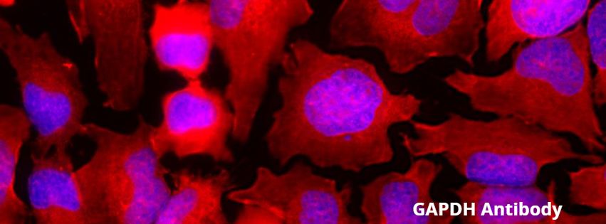
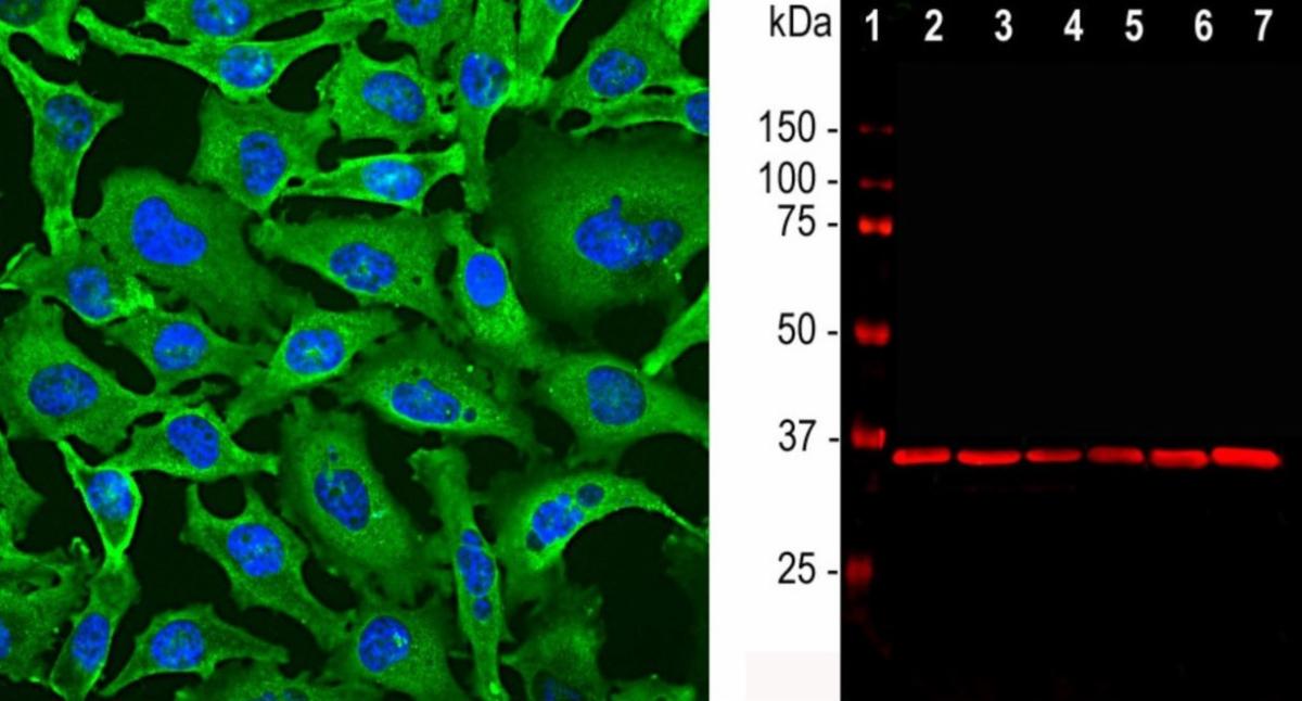
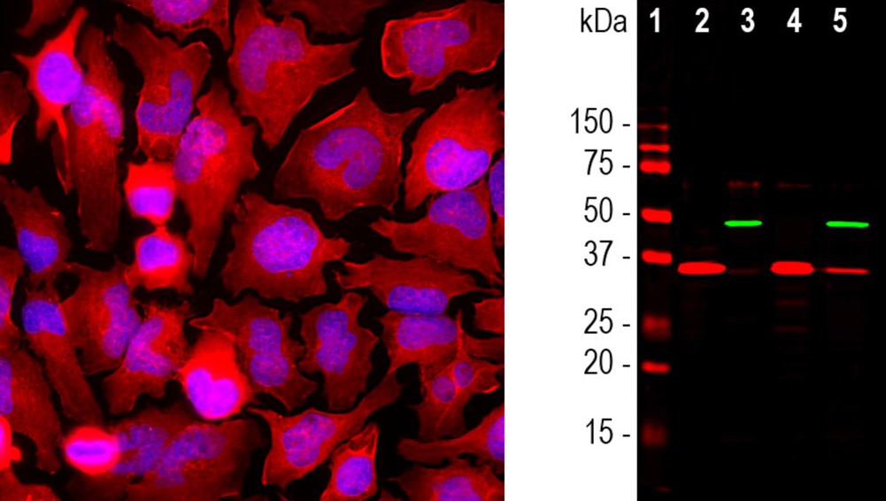
Biosensis offers two types of GAPDH antibodies: a mouse monoclonal antibody and a rabbit polyclonal antibody. These antibodies were specifically developed against highly purified pig GAPDH. They have become a widely used tool in research because of their reliable ability to detect GAPDH across multiple mammalian species, including humans, rats, and mice, and multiple applications including western blotting, immunocytochemistry, and immunohistochemistry, including paraffin-embedded tissues, They recognize an epitope within the peptide sequence corresponding to amino acids 254-273 of the human GAPDH protein, ensuring precise detection and quantification in various experiments.


Our antibodies are recognized for their quality and have been referenced in leading journals, including Neuroscience, Neurochemistry, Nutritional Journal, Journal of Neuroscience Research, International Journal of Molecular Sciences, Neuroscience Disability, and Aging Cell.

GAPDH,Mouse Monoclonal Antibody

(M-1376-250)

Above, leftStaining of GAPDH in HeLa cells with mouse anti-GAPDH antibody (green,

M-1376-250) by Immunocytochemistry. 


Above right: Western blot analysis of cell lysates probed with mouse antibody to GAPDH (red, lanes 2-7).

The GAPDH antibody reveals a single band at about 37 kDa in all cell lines.

GAPDH, Rabbit Polyclonal Antibody

(R-1701-100)

Above, left: HeLa cells stained with rabbit antibody to GAPDH (red, R-1701-100) by Immunocytochemistry. 


Above, right: Western blot analysis of cell cytosolic- or nuclear-enriched fractions, using rabbit antibody to GAPDH (red, lanes 2-5).

A strong band at 37 kDa corresponds to GAPDH protein, mainly detected in the cytosolic fractions. 

Enhance your research with our precise and versatile GAPDH antibodies

Discover our GAPDH Antibodies here

Biosensis is a company that is passionate about Science

Email  LinkedIn  Web