GFAP antibodies: don't mess around!

Dear Researcher,


Glial fibrillary acidic protein (GFAP, or class-III intermediate filament protein) is a classical biomarker expressed in astrocytes in the CNS, non-myelinating Schwann cells in the PNS, enteric glial cells and ependymal cells. A new study has identified that some GFAP polyclonal antibodies cross-react with desmin, which is closely related protein.

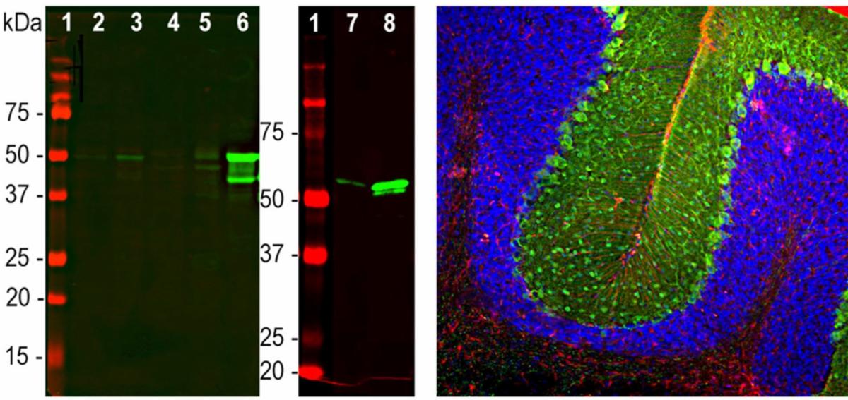
Left: Western blot analysis of GFAP expression in tissue homogenates. Primary antibody (M-1827green).

[1] protein standard, [2] rat brain, [3] rat spinal cord, [4] mouse brain, [5] mouse spinal cord, [6] pig brain, [7] rat recombinant GFAP, [8] human recombinant GFAP.

Right: IHC analysis of GFAP expression in an adult rat cerebellum section. Primary antibodies: mouse anti-GFAP

(M-1827, red), chicken anti-Parvalbumin (green) and DAPI nuclear stain (blue).

Pay attention to your GFAP antibody!


Gunther et al. (2021) investigated the distribution of GFAP outside of the brain and found positive staining in several organs due to cross-reactivity with desmin. They identified by WB and IHC analyses (see Table 1) that some GFAP antibodies cross-react with desmin whereas others do not.

Biosensis offers a range of antibodies including these two mouse monoclonals

(M-1375 and M-1827). Our GFAP antibodies are well cited in high-impact journals.

Table 1: Summary of WB and IHC analyses

For easy online ordering, head to our new website, or contact our friendly sales team at sales@biosensis.com.


For more information about our products email biospeak@biosensis.com.


Best wishes for your research,


The Biosensis Team


Twitter  Linkedin  
Biosensis Pty Ltd

Our mailing address is: