|
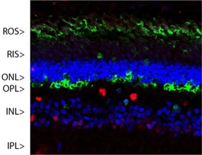
Left: Immunohistochemical analysis of arrestin-1 distribution in pig retina. Arrestin-1
(M-1796-100; green) is most abundant in the outer segments (OS) and inner surface of the outer nuclear layer (ONL). Other layers like IS, OPL, INL, and IPL display arrestin presence. The red stain shows staining for FOX2 (C-2127-100). Blue staining indicates nuclear DNA.
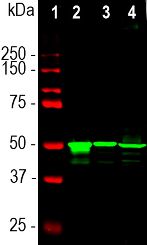
Right: Western blot analysis of retina lysates from different species using mouse mAb to arrestin-1, M-1796-100 in green: [1] protein standard (red), [2] rat [3] mouse and [4] cow retina lysates. The M-1796-100 antibody detects arrestin-1, running at about 48kDa.
|