Explore the Vital Role of Vimentin in Neuroscience with Biosensis Antibodies

Vimentin plays a crucial role in neural development, injury, and disease. It orchestrates cell migration and supports neurite outgrowth and axonal guidance. It also influences neural plasticity and cellular interactions with the extracellular matrix. Vimentin is a biomarker for diagnosing and monitoring neurodegenerative diseases. Its breakdown products can indicate brain injury severity, and increased levels correlate with disease progression in Alzheimer’s and multiple sclerosis. Biosensis offers research vimentin antibodies to facilitate vimentin research that will help contribute to our understanding of its roles in both healthy an diseased neurological conditions.

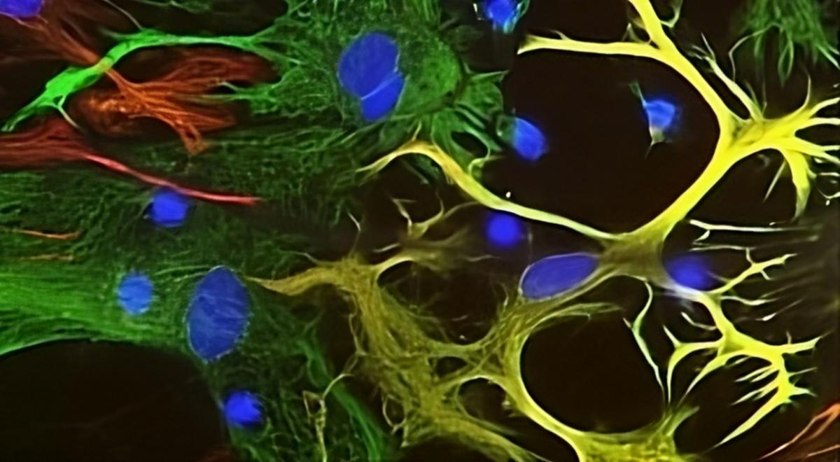
Title figure: View of mixed neuron/glial cultures stained with Chicken polyclonal antibody to Vimentin C-1409-50 (green) and Rabbit polyclonal antibody to Glial Fibrillary Acidic Protein R-1374-50 (red). Vimentin is expressed alone in fibroblastic and endothelial cells, which are the flattened cells in the middle of the image that appear green. Astrocytes may express primarily Glial Fibrillary Acidic Protein (GFAP), or GFAP and vimentin, and so appear red (GFAP only) or golden yellow (GFAP and Vimentin). In cells expressing GFAP and vimentin, the two proteins assemble to produce heteropolymer filaments.

Biosensis manufactures vimentin antibodies that are precisely engineered to deliver reliable results. These antibodies are essential tools for researchers who want to explore the complex roles of vimentin in neurobiology. Our antibodies exhibit high specificity and excellent sensitivity and are ideal for common applications such as Western Blotting and IHC. They are reactive with human, mouse, and rat cells and tissues and are the perfect tool for studying the role of vimentin protein in neural development, injury response, and disease progression.

C-1409-50

Left: Analysis of vimentin expression in HeLa cells by Immunocytochemistry. Cells were stained with chicken antibody to vimentin (C-1409-50, green), and co-stained with a mouse anti-actin antibody (M-1646-100, red). Blue: DAPI nuclear stain.

Right: Western blot analysis of tissue and cell lysates using chicken antibody to Vimentin (C-1409-50, red).

M-2121-100

Left: Immunofluorescent analysis reveals vimentin expression in rat cortical neuron-glial cell culture. The section was stained with mouse anti-Vimentin antibody (M-2121-100, green) and co-stained with rabbit polyclonal antibody to GFAP (R-1374-50, red), with DAPI staining indicating nuclear DNA (blue).

Right: Western blot analysis of whole brain tissue lysates using (M-2121-100, green) reveals a band at approximately 50 kDa.

R-1699-100

Left: Immunocytochemistry reveals vimentin expression in HeLa cells using a rabbit antibody to vimentin (R-1699-100, green). Co-staining with an antibody to actin is represented in red (M-1646-100), while DAPI nuclear stain is in blue. The vimentin antibody effectively highlights the 10 nm or intermediate filament network of the cytoskeleton.

Right: Western blot analysis of whole cell lysates utilizing the rabbit antibody to vimentin (R-1699-100, green) to show the prominent band at approximately 55 kDa corresponds to the vimentin protein.

Elevate your research with our Vimentin Antibodies

Explore more

Biosensis is a company that is passionate about Science