SHARE:  

Calbindin: Key Marker for Neuronal Study

Explore Neuronal Diversity with Our Calbindin Antibodies

Calbindin is a calcium-binding protein highly expressed in inhibitory GABAergic interneurons and some excitatory neurons. It aids in calcium buffering and protection and is used as a neuronal marker in neuroscience. Calbindin staining is essential for studying neuronal subtypes and brain function.

Explore Neuronal Diversity with Our Calbindin Antibodies

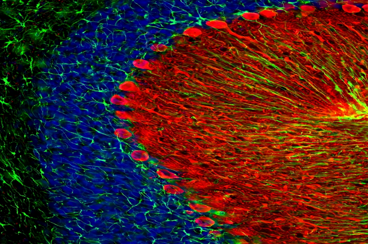
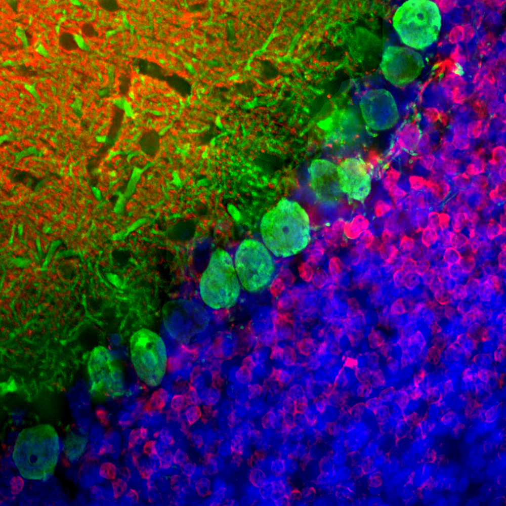
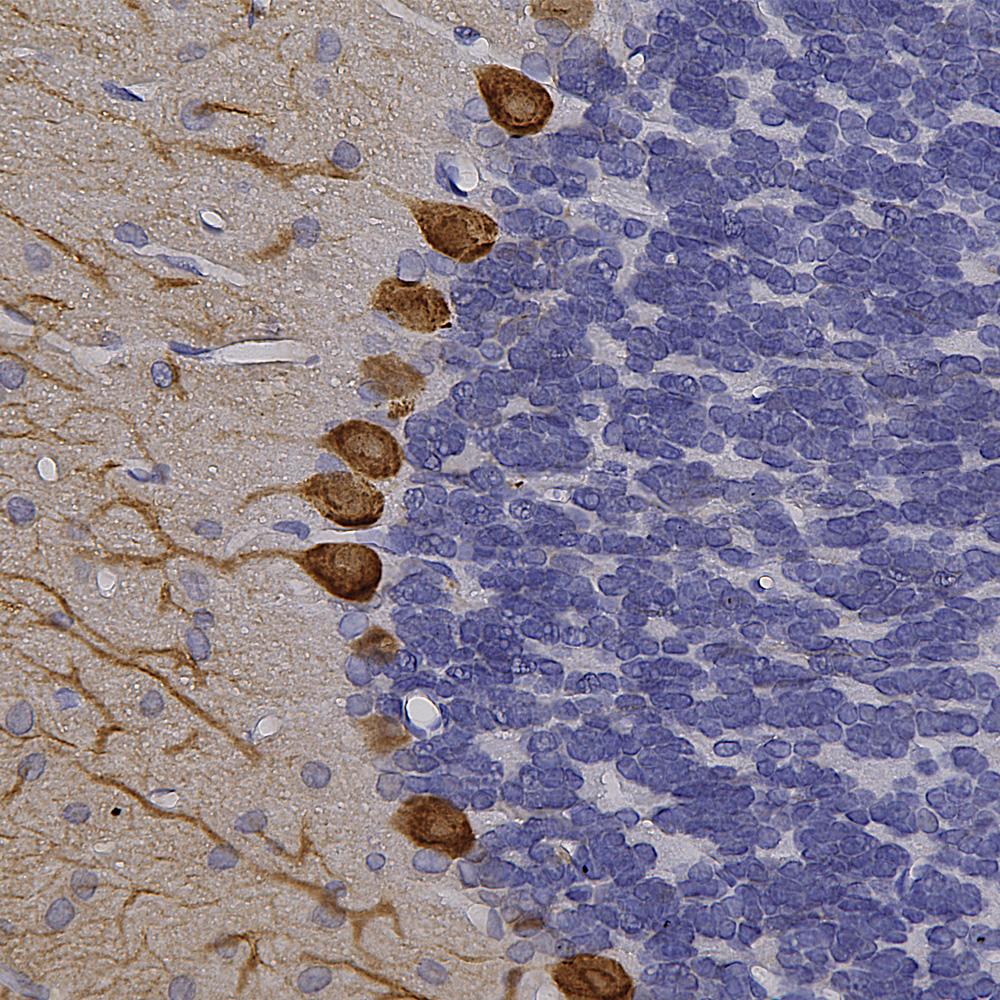
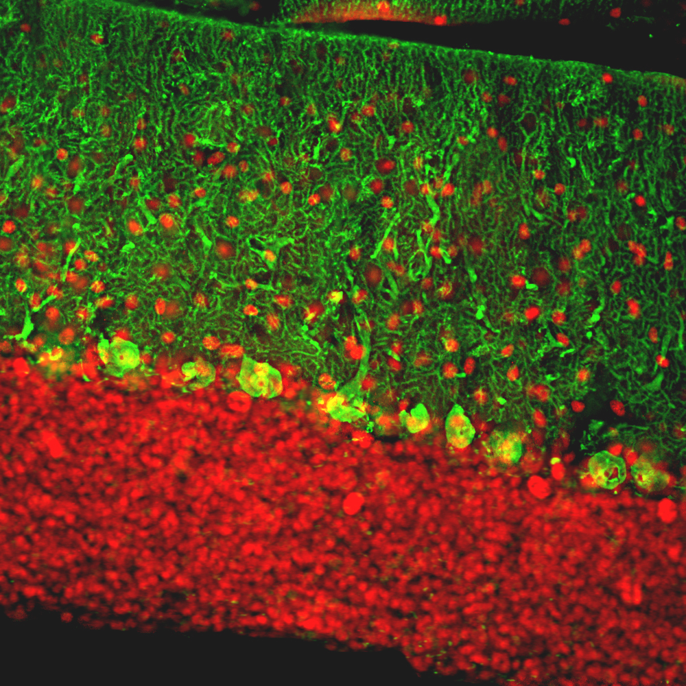
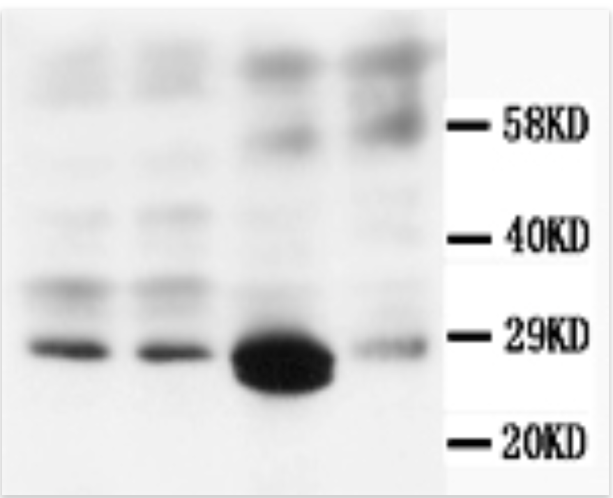
Biosensis developed chicken and mouse antibodies against full-length recombinant human calbindin, which was expressed and purified from E.coli. Our research has shown that these antibodies specifically bind to calbindin in western blots and tissue sections without cross-reacting with the related proteins calretinin and parvalbumin. This specificity makes them ideal for identifying calbindin and subclassifying cortical GABAergic neurons. Our antibodies are effective for western blotting, immunofluorescence (IF), immunocytochemistry (ICC), and immunohistochemistry (IHC), and they are reactive against human, mouse, rat, and bovine samples, depending on the selected catalog number.

The immunofluorescence image of rat cerebellum shows the staining of mouse anti-calbindin (M-1797-100, green) and rabbit anti-calretinin (R-1042-100, red). DAPI (blue) marks nuclear DNA. Calbindin highlights Purkinje cells, while calretinin stains granule cells and their processes.

DAB chromogenic immunostaining of a formalin-fixed paraffin-embedded rat cerebellum section with Biosensis chicken polyclonal antibody to calbindin, C-1798-50, dilution 1:10,000 (brown), with blue hematoxylin counterstain. This antibody strongly stains Purkinje cell perikarya and dendrites nicely in formalin-fixed paraffin-embedded tissues.

The immunofluorescence image of rat cerebellum shows the staining of calbindin chicken polyclonal

(C-1798-50, green) and rabbit anti-MeCP2

(M-809-100, red). DAPI (blue) marks nuclear DNA. Calbindin highlights Purkinje cell dendrites and perikarya, while MeCP2 labels neuronal nuclei.

Western blot of mouse monoclonal antibody to Calbindin-D (M-976-100) using rat brain lysate (lanes 1 and 2), rat kidney lysate (lane 3), and rat lung tissue lysate (lane 4) at a concentration of 0.5 µg/mL. Secondary antibody HRP conjugated goat anti-mouse IgG at 1:3,000-10,000.

Elevate your research with our Calbindin antibodies. Learn more at the links below:

Biosensis is a company that is passionate about Science