|
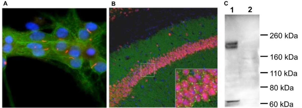
A: Immunofluorescence staining of neuronal cilia in rat neuron-glial cultures using
anti-ACIII polyclonal antibody (R-1687-100; red). Co-stained with neuronal alpha-II spectrin (M-1575-100; green) and DNA (blue).
B: Immunohistochemistry of mouse brain sections using anti-ACIII polyclonal antibody (R-1687-100; green) antibody reveals specific cilia labeling near pyramidal neurons in the CA1 hippocampus region, while other brain areas remain unlabeled. Co-stained with anti-Fox3/NeuN (M-377-100; red) and DAPI for cell nuclei (blue).
C: Western blot analysis of rat olfactory epithelium (Lane 1) and frontal cortex (Lane 2) using anti-ACIII polyclonal antibody (R-1687-100). It detects a prominent 200 kDa protein band in cilia-rich olfactory epithelium, while the frontal cortex, with fewer cilia and lighter glycosylation, shows a less prominent 160 kDa band.
|